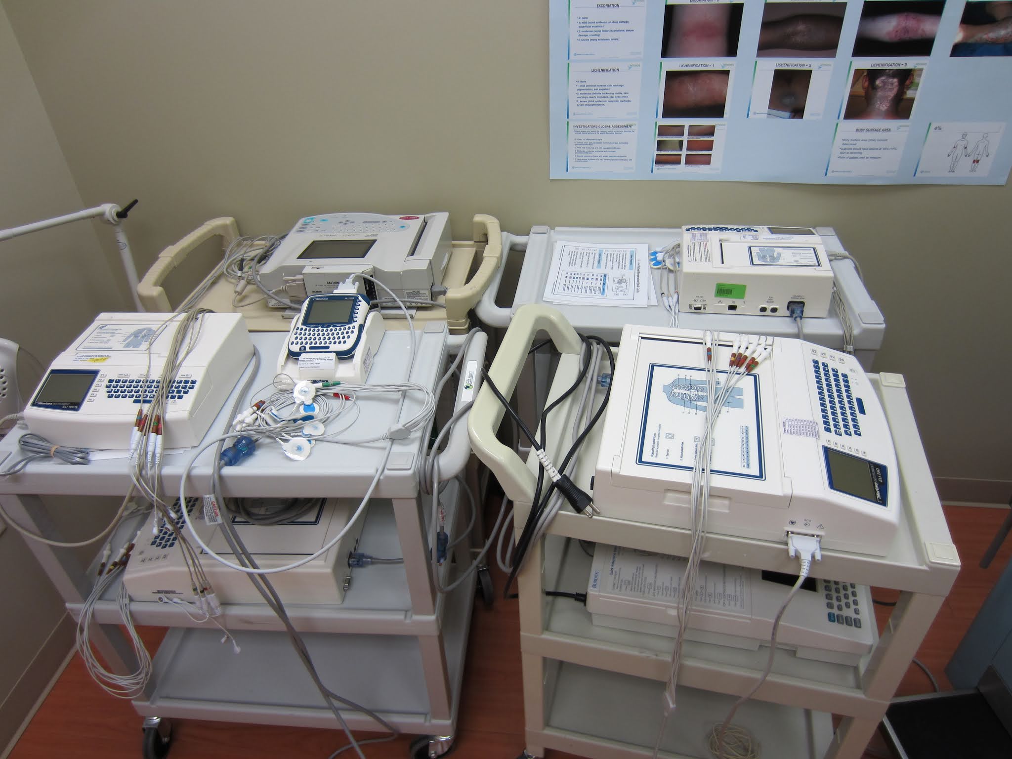
What is The ECG Standardization ।। Clarify The Reason For Normalization of The ECG ।। Normalization And Change of The Electrocardiography Meaning

An electrocardiogram, or ECG, is a perusing evaluating the extent and course of the electrical flows of the heart and estimating the depolarization and repolarization of the cardiovascular muscle cells
It's anything but an ECG that is recorded precisely.
ECG cathode situation is normalized, considering the account of a precise follows - yet additionally guaranteeing similarity between records taken at various occasions.
Helpless cathode arrangement can bring about mixed-up translation, which may then prompt conceivable misdiagnosis, patient botch, or unseemly strategies. Deviation of lead arrangement even by 20-25mm from the right position can make clinically critical changes on the ECG, including changes to the ST-section.
Patient factors like breath, position, smoking, ongoing dietary admission, and weight may likewise add to the exactness of an ECG perusing.
Not just guarantee that the terminals are set as per the normalized 'rules', yet additionally, that the patient is arranged accurately for the technique, both actually and mentally.
Setting up a Patient for an ECG
Similarly, as with all methodology, you should acquire educated assent from the patient by clarifying the reason for the strategy, portraying the actual system, and getting agree to continue. Keep up with great contamination control practice by washing your hands preceding patient contact.
Skin arrangement is significant. On the off chance that the patient's skin is grimy, clean with cleanser and water, and dry. On the off chance that the skin is slick or the patient applied any creams or moisturizers, utilize a liquor wipe to clean every terminal position site.
Some ECG machines may likewise give a 'difficult time' either independently or on the cathodes, which can be utilized to rub on the skin to expand terminal adherence. Care ought to be taken not to cause scraped areas.
Patients with chest hair ought to have hair at the cathode situation destinations eliminated with a hair trimmer.
Where conceivable, place the patient in a prostrate or semi-supine situation with their legs and arms uncrossed. On the off chance that this is absurd or is awkward for the patient, it is satisfactory to record the ECG in another position.
The patient should be totally loose. Guarantee the climate is at a serenely warm temperature. This will keep strong pressure or developments from creating curio on the ECG recording. Guarantee the patient's security and poise: for example by shutting the room entryway or closing around the draperies.
12-Lead ECG Placement

The patient's chest and every one of the four appendages ought to be presented to apply the ECG anodes effectively.
There are various strategies for recognizing the right tourist spots for ECG terminal arrangement, the two most normal being the 'Point of Louis' Method and the 'Clavicular' Method.
ECG anodes are shading coded, and each is recognized by a particular code that alludes to its expected situation. There are two coding frameworks as of now being used:
American Heart Association (AHA) framework
Worldwide Electrotechnical Commission (IEC) framework.
The two frameworks are portrayed in the table underneath.
Code (AHA) Code (IEC) Location Color (AHA) Colour (IEC)
V1 C1 fourth intercostal space at the right sternal border Brown/red White/red
V2 C2 fourth intercostal space at the left sternal border Brown/yellow White/yellow
V3 C3 Halfway between drives V2 and V4 Brown/green White/green
V4 C4 Fifth intercostal space in the midclavicular line Brown/blue White/brown
V5 C5 Left foremost axillary line on a similar even plane as V4 Brown/orange White/dark
V6 C6 Left midaxillary line on a similar even plane as V4 and V5 Brown/purple White/purple
RA R Right arm (internal wrist) White Red
LA L Left arm (internal wrist) Black Yellow
RL N Right leg (internal ankle) Green Black
LL F Left leg (internal ankle) Red Green
(Adjusted from Crawford and Doherty 2020; Jevon 2010; Cables and Sensors 2021)
Precordial Lead Placement
Note: The accompanying aide utilizes the AHA framework.
To discover these accurately, the 'Point of Louis' Method can be utilized:
To find the space for V1; find the sternal score (Angle of Louis) at the subsequent rib and feel down the sternal line until the fourth intercostal space is found. V1 is set to one side of the sternal boundary, and V2 is set at the left of the sternal line.
Then, V4 ought to be set before V3. V4 ought to be set in the fifth intercostal space in the midclavicular line (as though defining a boundary downwards from the focal point of the patient's clavicle).
V3 is put straightforwardly somewhere in the range of V2 and V4.
V5 is put straightforwardly somewhere in the range of V4 and V6.
V6 is put over the fifth intercostal space at the mid-axillary line (as though defining a boundary down from the armpit).
V4-V6 should arrange evenly along the fifth intercostal space.
#chelseabibi777
#healthtec


0 Comments